X-RAY DIAGNOSTICS AT MVZ RESTART-ORTHO
X-ray diagnostics are among the indispensable procedures in modern orthopedics. They enable a quick, reliable, and structured assessment of bony structures, joint axes, joint space ratios, and degenerative changes. As an initial examination, it provides essential foundations for further diagnostics and therapy planning for acute and chronic complaints of the musculoskeletal system.
At MVZ Restart-Ortho, X-ray diagnostics are fully integrated into the interdisciplinary treatment process. Modern digital technology, standardized implementation, and direct medical evaluation ensure precise images that flow directly into medical decision-making.
- 1. When is an X-ray examination indicated in orthopedics?
- 2. What advantages does X-ray offer compared to other imaging methods?
- 3. Which body regions can be examined using X-ray?
- 4. How does an X-ray examination work at MVZ Restart-Ortho?
- 5. What role does X-ray diagnostics play in therapy planning?
- 6. What advantages does the implementation in our own center offer?
- 7. Frequently asked questions about X-ray diagnostics
- 8. Frequently asked questions about aftercare and follow-up with X-ray
1. When is an X-ray examination indicated in orthopedics?
An X-ray examination is used in almost all areas of orthopedic diagnostics. It is particularly indicated for acute pain after trauma, restricted movement, chronic joint complaints, malpositions, and suspected degenerative or inflammatory changes. In the area of the knee joint, X-rays allow a precise assessment of the joint space width, the bony structures of the femur, tibia, and patella, and the position of the joint partners in relation to each other.
For shoulder complaints, X-ray diagnostics provide important information on calcium deposits, arthrotic remodeling, or structural changes to the humeral head, glenoid, or acromion.. In hip diagnostics, X-rays can be used to display dysplasias, signs of arthrosis, or impingement-constellations. X-rays also play a central role in complaints of the spine, for example in the assessment of poor posture, degenerative changes in the vertebral bodies, height reduction of intervertebral discs, or instabilities.
If there is a suspicion of fractures, malpositions, or joint wear, X-rays are often the first and often sufficiently clarifying measure. In addition, it is indispensable in orthopedic aftercare, for example for follow-up checks after operations or conservative treatments.

2. What advantages does X-ray offer compared to other imaging methods?
Conventional X-ray is a fast, cost-effective method that has been tried and tested for decades for imaging the skeletal system. It is almost universally available, has a very low radiation exposure thanks to modern digital X-ray technology, and is ideally suited for assessing bony structures, joint spaces, axial ratios, and implant positions.. In contrast to magnetic resonance imaging, which primarily images soft tissues, cartilage, and ligaments, X-ray diagnostics focuses on hard tissue structures such as bones, joint contours, and bony remodeling. For numerous questions, including arthrosis, osteoporosis, fractures, postoperative follow-up examinations, or structural malpositions, X-rays are not only sufficient, but often the means of first choice. In addition, the standardized implementation enables reliable comparability over longer periods of time, for example when observing the course of the disease or checking the stability of surgically treated areas.
3. Which body regions can be examined using X-ray?
X-ray diagnostics can be used in all major joint areas as well as on the spine and offers a high degree of informative value in orthopedics. At the knee joint, it allows the precise assessment of the joint space ratios, the patella height and -guidance as well as the bony alignment of the tibia and femur, which also allows degenerative changes such as meniscus wear or axial malpositions to be recognized. On the shoulder, X-rays can be used to display calcium deposits, joint space narrowing, osteophytes, or joint instabilities, as well as degenerative changes in the acromioclavicular joint or bony impingement syndromes.
In the hip, X-rays provide reliable information about coxarthrosis, deformities, or femoroacetabular impingement-constellations. On the spine, height reductions, changes in the vertebral bodies, scoliosis, or spondylolistheses can be documented. Even smaller joints such as the wrist, ankle, or elbow can be displayed in detail with targeted images. The diagnostic benefit always depends on the clinical question, but X-rays have an outstanding significance in orthopedics as a basic examination for complaints of the musculoskeletal system.
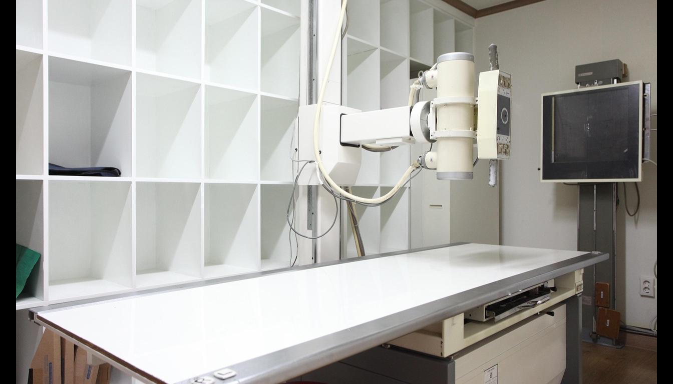
4. How does an X-ray examination work at MVZ Restart-Ortho?
An X-ray examination at MVZ Restart-Ortho is carried out in a standardized manner, quickly, and always in close coordination with the specific orthopedic question. After a brief preliminary discussion and the targeted positioning of the body, the image is taken with a modern digital X-ray machine. Depending on the region to be examined, for example in the case of a full-leg image, a clarification of the spine, or X-ray diagnostics on the knee, the examination is carried out while standing, while for certain injuries such as a shoulder dislocation or a patellar dislocation, it is carried out while lying down. Thanks to the digital technology, the images are immediately available and can be evaluated by a doctor immediately.
In addition to the high image quality, the clinical relevance of the images is in the foreground, so that the imaging fits exactly to the described complaints – be it to clarify a cruciate ligament rupture, for meniscus tear treatment, for control after an operation or for the assessment of degenerative changes in the hip, shoulder or spine. The data obtained flows directly into medical decision-making. As a rule, the entire process, including preparation, recording, and medical discussion, only takes a few minutes, which enables quick decisions, such as the immediate initiation of therapy, especially in orthopedic emergencies.
5. What role does X-ray diagnostics play in therapy planning?
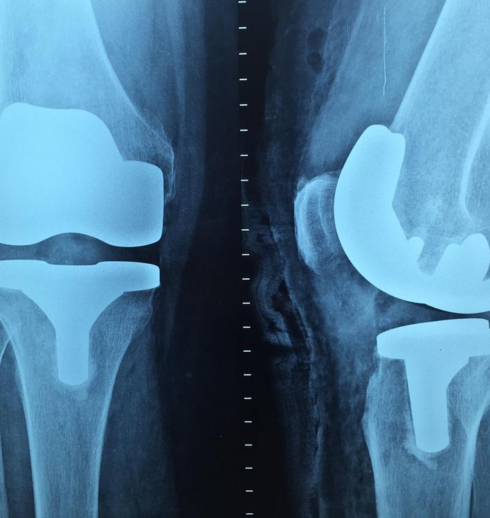
X-ray diagnostics often form the basis of a well-founded orthopedic therapy decision. If joint wear is suspected, it shows not only the extent but also the exact localization of degenerative changes and thus provides valuable information as to whether conservative therapy – for example in combination with autologous blood therapy on the knee – makes sense or whether an operative measure is necessary. In preoperative planning, for example in the case of arthrotic knee joints, X-rays can be used to precisely assess whether a joint-preserving procedure is possible or whether a joint replacement is the better option.
X-rays also offer a simple and safe way to monitor the healing process, check the position of screws, plates, or implants, and identify possible complications early on in the aftercare after operations – such as osteosyntheses, conversion osteotomies, or the use of endoprostheses. In conservative therapies, it is used regularly to document the course of diseases such as arthrosis, osteoporosis, or chronic malpositions and to adapt the treatment individually.
6. What advantages does the implementation in our own center offer?
The greatest advantage of X-ray diagnostics directly at MVZ Restart-Ortho lies in the immediate availability of the examination and the continuous quality control from image acquisition to evaluation. Since no referral to external facilities is required, diagnoses can be made immediately in many cases and therapies can be initiated directly. This is particularly advantageous in the case of acute injuries such as a cruciate ligament rupture, a shoulder dislocation, or a patellar dislocation. All image data is stored centrally, which means that later follow-up checks with the initial images are easily possible – without loss of information or media breaks.
The close interlinking of X-ray, medical evaluation, and therapy decision guarantees that exactly the question relevant to the patient is answered. Even complex findings, such as combined ligament and cartilage damage, implant loosening, or axial deviations, can be clarified efficiently and from a single source. For patients, this means not only a significant saving of time, but also a high degree of transparency in the course of treatment and continuous care by an experienced orthopedic team.
7. Frequently asked questions about X-ray diagnostics
Is X-ray dangerous? How high is the radiation exposure?
The radiation exposure from modern digital X-ray images is very low and usually well below the natural background radiation that people are exposed to, for example during air travel or stays in the mountains. Through targeted dose control, radiation-reduced technology, and precise focusing of the images, only the required area is examined—whether in X-ray diagnostics of the knee after a cruciate ligament tear, the shoulder in cases of suspected dislocation, or the spine in cases of chronic complaints. The medical necessity of each image is carefully evaluated in advance. In weighing the benefits and risks, the diagnostic value of an X-ray examination generally far outweighs the minimal radiation risk—particularly in structural conditions, acute injuries, or orthopedic emergencies.
Why is an X-ray often performed first instead of an MRI?
X-ray is a fast, cost-efficient, and well-established procedure that has held a central role in orthopedic primary care for decades. It provides reliable information on bone structure, joint alignment, signs of osteoarthritis, or fractures—often sufficient for an initial therapeutic decision. In many cases, such as the assessment of a meniscus tear, a patellar dislocation, or advanced joint degeneration, X-ray diagnostics already offer a clear basis for further treatment. An MRI examination is used additionally when soft tissue injuries such as ligament tears, meniscus, or tendon lesions are suspected, or when symptoms persist despite an unremarkable X-ray image. The two methods complement each other, with X-ray serving as the first diagnostic step in most orthopedic cases.
8. Frequently asked questions about aftercare and follow-up with X-ray
How often may or should X-rays be taken – for example in cases of chronic complaints?
The frequency of X-ray examinations always depends on medical necessity and the individual clinical condition. In chronic diseases such as osteoarthritis or malalignments, a check-up every six to twelve months is usually sufficient. In cases of acute complaints, after injuries such as a cruciate ligament tear, a non-surgically treated meniscus tear, a shoulder dislocation, or a patellar dislocation, closer monitoring may be required—especially after surgical procedures or when adjusting conservative therapy. Throughout this process, strict attention is paid to minimizing radiation exposure. Every image is medically justified and planned to provide maximum diagnostic value while keeping risk to an absolute minimum.
Can the progression of a disease be reliably tracked using X-rays?
X-ray imaging provides a reliable way to document structural changes over time. For example, it allows an objective assessment of the progression of osteoarthritis, the healing process after a fracture, the ingrowth of a prosthesis, or the correction of a malalignment. Even after joint-preserving procedures or additional treatments such as autologous blood therapy of the knee, imaging can deliver crucial insights into therapeutic success. Thanks to digital storage within the MVZ’s internal system, direct image comparisons are possible without difficulty—even after many years. This continuous follow-up supports precise, individualized treatment planning and offers patients a high level of transparency and safety.