MRI DIAGNOSTICS AT MVZ RESTART-ORTHO
MRI diagnostics at MVZ Restart-Ortho enables precise and radiation-free imaging of joints, soft tissues, and internal structures. Using state-of-the-art technology and specialized expertise, it makes a significant contribution to rapid and well-founded diagnosis.
- 1. When is an MRI useful, e.g., for leg axis misalignments?
- 2. How informative is MRI compared to X-ray?
- 3. What is the procedure for an MRI examination at the center?
- 4. Which structures can be assessed exactly with MRI?
- 5. What role does MRI play in therapy planning?
- 6. What are the advantages of “in-house” MRI?
- 7. What alternatives to MRI are there – and what are their limitations?
- 8. What is the significance of MRI in follow-up monitoring?
1. When is an MRI useful, e.g., for leg axis misalignments?
MRI is particularly useful when there are complaints in the area of the knee joint, the cause of which goes beyond simple X-ray diagnostics. MRI provides important information, especially in the case of unclear pain that occurs during exertion, swelling, suspected meniscus injuries, or cartilage damage. MRI can also make a decisive contribution to therapy planning in the case of progressive misalignment of the leg axis or in the case of therapy-resistant complaints despite conservative measures.
MRI is essential, especially in patients in whom operative axis correction (e.g., osteotomy) is being considered: It is used to assess the condition of the articular cartilage, to depict possible structural damage, and thereby to determine the appropriate surgical indication and strategy.

2. How informative is MRI compared to X-ray?
The conventional X-ray – especially the so-called “full-leg standing image” – is essential for assessing the mechanical leg axis and load distribution when standing. It shows bony misalignments, leg length differences, or pathological axis deviations. However, its informative value is limited when it comes to assessing soft tissues, cartilage, or inflammation.
This is where MRI offers clear advantages: It can image the smallest structural changes long before they become visible on X-rays. Cartilage defects, meniscus degeneration, joint effusions, or cruciate ligament damage can be reliably detected. In combination, this results in a complete picture – structurally, functionally, and prognostically.
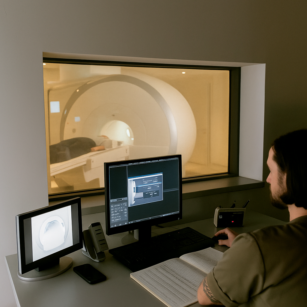
3. What is the procedure for an MRI examination at the center?
At MVZ Restart-Ortho, the MRI examination is fully integrated into the care structure. This means that after the clinical examination and the medical consultation, the MRI can often be performed within a few days – sometimes even on the same day. There are no long waiting times for external appointments. The coordination between orthopedics and radiology also takes place in-house and seamlessly.
Before the examination, the exact question is coordinated with the radiology department so that targeted sectional images can be taken. The examination itself lasts between 20 and 30 minutes, is non-invasive, and can be carried out without any problems for most people. Since MRI works without ionizing radiation, it is also suitable for follow-up monitoring or for younger patients.
After the examination, the findings are prepared by experienced radiologists and are usually available to the treating orthopedists very quickly. On this basis, further therapy is planned – individually, well-founded, and evidence-based.
4. Which structures can be assessed exactly with MRI?
Magnetic resonance imaging allows an extremely differentiated representation of all structures near the joint and is therefore particularly suitable for visualizing complex damage or changes in the area of the knee joint. The assessment of the articular cartilage is particularly relevant, as its condition is decisive for therapeutic planning. Early signs of incipient arthrosis – such as cartilage softening, superficial defects, or local thinning – can often be detected with MRI even when they are not yet visible on X-rays. Existing cartilage damage can also be precisely localized, assessed in terms of extent and severity, and thus classified for surgical planning.
In addition, MRI offers an exact representation of the menisci, which are often subject to degenerative changes or tears due to incorrect loading caused by leg axis misalignment. Band structures such as the anterior and posterior cruciate ligaments and the lateral ligaments are also clearly visible in the sectional image, so that both acute injuries and chronic instabilities can be reliably assessed. In addition to these intra-articular structures, the bony architecture, including possible edema, reactions of the subchondral bone, or accompanying cyst formation, can also be perfectly imaged. Soft tissue components such as bursae, tendon attachments, or free joint bodies can also be recorded. This comprehensive representation is particularly important when complex clinical pictures are present in which several structures may be affected at the same time.
5. What role does MRI play in therapy planning?
MRI is a central diagnostic tool for planning individually adapted therapy for leg axis misalignments. It provides not only structural information about the condition of the joint, but also functional information about stress conditions, signs of inflammation, or biomechanical compensations. MRI is of inestimable value, especially when deciding whether to continue with a conservative treatment approach or to perform a surgical intervention – such as an osteotomy. For example, a localized cartilage defect in combination with a varus axis deviation may indicate that operative relief of the medial joint component is necessary to protect the cartilage and slow the progression of degenerative processes.
Likewise, in the case of unclear or therapy-resistant complaints, MRI provides information on alternative causes of pain, such as a previously undiscovered meniscus lesion, irritation of the Hoffa fat pad, or synovial irritation, which cannot always be clearly assigned in the clinical findings. Imaging therefore supports not only the treatment decision itself, but also the differentiated clarification of prognosis, therapy alternatives, and potential risks. Above all, in the preoperative phase, it represents an indispensable addition to clinical and radiological diagnostics.
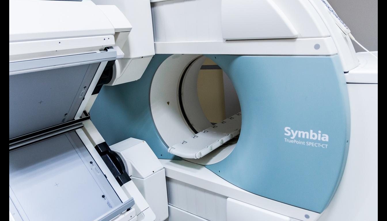
6. What are the advantages of “in-house” MRI?
The integration of MRI into the structures of MVZ Restart-Ortho creates a significant advantage for diagnostics and treatment. Since all the specialist disciplines involved – orthopedics, radiology, and therapy planning – work under one roof, processes can be coordinated efficiently and diagnostic gaps avoided. The organizational effort for patients is minimized, as referrals to external radiology departments are not necessary and appointments for MRI are made directly at the center. This not only leads to a significant saving of time, but also to seamless communication between the treating physicians involved.
The standardized reporting takes place in direct feedback with the orthopedic question, which guarantees a high level of diagnostic precision and clinical relevance. Thanks to direct access to the image data, a rapid initiation of further steps is possible – be it conservative treatment, an indication for surgery, or additional diagnostic clarification. In addition, internal coordination increases security in decision-making and ensures transparency and traceability in the therapeutic procedure. Ultimately, the entire treatment process benefits from the structural efficiency, the professional proximity, and the consistent patient management within the center.
7. What alternatives to MRI are there – and what are their limitations?
Was ist eine Umstellungsosteotomie und wann wird sie durchgeführt?
Eine Umstellungsosteotomie ist ein gelenkerhaltender operativer Eingriff, bei dem der Knochen gezielt durchtrennt und in einem veränderten Winkel wieder stabilisiert wird, um die mechanische Beinachse zu korrigieren. Ziel ist es, die Lastverteilung im Kniegelenk zu verändern, um überbeanspruchte Gelenkabschnitte – meist den inneren oder äußeren Anteil – zu entlasten. Die Operation kommt insbesondere dann zum Einsatz, wenn eine Beinachsenfehlstellung (z. B. O- oder X-Bein) zu Knorpelschäden oder arthrotischen Veränderungen in einem Teil des Gelenks führt, während andere Abschnitte noch gut erhalten sind. Eine rechtzeitig durchgeführte Umstellungsosteotomie kann so das Fortschreiten der Arthrose verzögern oder sogar stoppen und die Notwendigkeit eines künstlichen Gelenks um viele Jahre hinauszögern.
Wie läuft der Eingriff technisch ab und wie lange dauert er?
Die Operation erfolgt in der Regel minimalinvasiv oder offen chirurgisch unter Vollnarkose oder Spinalanästhesie. Dabei wird ein Teil des Schienbeins (Tibia) oder Oberschenkelknochens (Femur) präzise durchtrennt. Je nach Lage der Fehlstellung wird der Knochen entweder aufgeklappt (open wedge) oder ein Keil entfernt (closed wedge), um die gewünschte Achskorrektur zu erzielen. Die neue Position wird mit einer stabilen Titanplatte fixiert. Die Dauer des Eingriffs liegt meist zwischen 60 und 90 Minuten. Ein stationärer Aufenthalt von mehreren Tagen ist in der Regel erforderlich, die Belastbarkeit wird im Anschluss schrittweise aufgebaut. Die Operation ist technisch anspruchsvoll, erzielt jedoch bei guter Indikationsstellung ausgezeichnete funktionelle Ergebnisse.
8. What does the aftercare look like?
Wann darf nach der Operation wieder belastet werden?
Die Nachbehandlung nach einer Umstellungsosteotomie erfolgt stufenweise und orientiert sich am Heilungsverlauf des Knochens. In den ersten Tagen nach der Operation erfolgt eine Teilbelastung mit Unterarmgehstützen, um den operierten Bereich zu entlasten und eine stabile Knochenheilung zu gewährleisten. Je nach Ausmaß der Korrektur, Knochenqualität und individueller Belastbarkeit wird die Belastung in den folgenden Wochen sukzessive gesteigert – häufig begleitet durch Physiotherapie. Eine vollständige Belastung ist in der Regel nach 6 bis 8 Wochen möglich, sportliche Aktivitäten sind meist nach 3 bis 6 Monaten wieder erlaubt. Kontrolluntersuchungen und bildgebende Verlaufskontrollen begleiten diesen Prozess und dienen der Anpassung des Rehabilitationsplans.
Welche Maßnahmen unterstützen eine optimale Heilung nach dem Eingriff?
Eine konsequente Nachbehandlung ist entscheidend für den langfristigen Erfolg der Operation. Dazu gehört neben der ärztlich begleiteten Mobilisation vor allem eine physiotherapeutisch angeleitete Bewegungstherapie, die auf Muskelaufbau, Gelenkstabilität und Beweglichkeit abzielt. Auch das Einhalten der verordneten Belastungsgrenzen, die regelmäßige Lymphdrainage zur Schwellungsreduktion und gegebenenfalls eine Thromboseprophylaxe in der Frühphase sind wichtige Bestandteile der Therapie. Je nach individuellem Verlauf kann zusätzlich eine osteoanabole Medikation oder Vitamin-D-Substitution sinnvoll sein, um die Knochenheilung zu unterstützen. In den meisten Fällen ist die knöcherne Konsolidierung nach 3 bis 4 Monaten abgeschlossen, wobei die vollständige Funktionswiederherstellung weitere Zeit in Anspruch nehmen kann.