OPTIMAL TREATMENT OF LEG AXIS MISALIGNMENTS - OVERVIEW
Leg axis malalignments lead to asymmetrical joint loading and promote degenerative changes in the knee. The aim of therapy is to preserve the joint by balancing the load.
- 1. What is a leg axis malalignment?
- 2. How does a leg axis malalignment develop?
- 3. What complaints occur with leg axis malalignments?
- 4. How is a leg axis malalignment diagnosed?
- 5. What is the optimal therapy for leg axis malalignments?
- 6. How is an osteotomy performed?
- 7. Frequently asked questions about osteotomy
- 8. What does the aftercare look like?
1. What is a leg axis malalignment?
A leg axis misalignment is defined as a deviation from the physiological, i.e. natural, straight alignment of the leg axis. Normally, the leg axis runs from the hip via the knee joint to the ankle joint almost straight. A deviation can be inwards, as a knock-knee or valgus position, or outwards, as a bow-leg or varus position.
Such changes lead to an uneven distribution of pressure in the knee joint, which mechanically overloads individual joint sections. In the long term, this can promote the development of cartilage damage, meniscus injuries or osteoarthritis. Rotational misalignments, for example after bone fractures, as well as unstable positions of the kneecap can also cause biomechanical overloads. It is particularly problematic if a misalignment remains unnoticed for a long period of time.
The aim of every therapeutic measure is therefore to restore the most physiological load on the joint possible, to reduce pain and to maintain joint function in the long term, if possible before irreversible damage occurs.
2. How does a leg axis malalignment develop?
Leg axis misalignments can be congenital or acquired during the course of life. Often there is a combination of several influencing factors. Congenital or developmental misalignments often occur in childhood or adolescence due to disturbances in bone growth.
If the femur or tibia grow at different rates or in the wrong direction, this can cause a deviation of the axis. A genetic predisposition can increase the risk, especially if there is already a knock-knee or bow-leg position in the family. Slight deviations often correct themselves during growth, but more pronounced or permanent misalignments should be clarified by a doctor.
Acquired misalignments occur more frequently in adulthood and arise, for example, after bone fractures that heal in a misaligned position, especially in the area of the femoral neck, the tibia or the ankle joint.
Long-term one-sided stress at work or excessive sporting activity can also change the leg axis. Repeated patellar dislocations with unstable joint guidance can also cause an axis deviation in the long term. Often such changes initially go unnoticed and are only recognised by increasing pain or restricted movement.
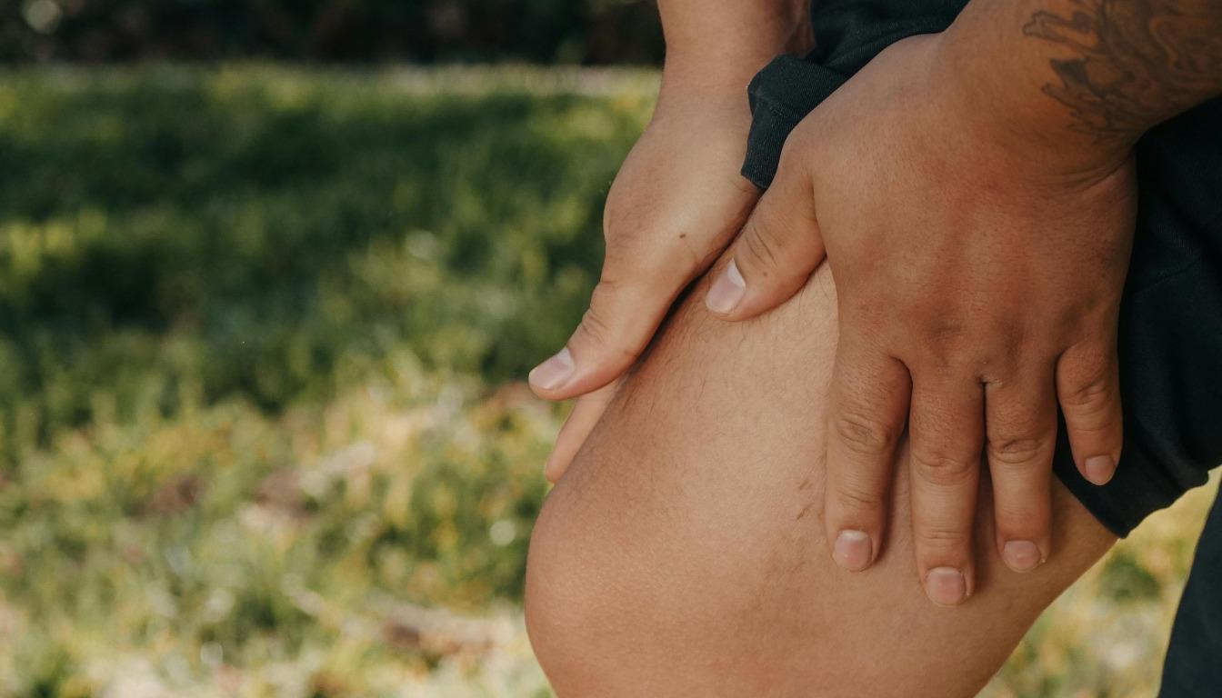
3. What complaints occur with leg axis malalignments?
The symptoms usually develop insidiously and are unspecific at the beginning. Frequently, there is an asymmetrical load on the knee joint, which is particularly noticeable when walking, standing or doing sports for longer periods of time. Load-dependent pain usually occurs on the more stressed side of the knee, a lso outside with a knock-knee position or inside with a bow-leg position. Swelling may occur in the course, especially after intense stress or longer walks. Many affected people report a subjective feeling of instability as well as restricted movement.
Recurring irritation and inflammation in the joint area are common, and in advanced stages osteoarthritis can occur. Untreated leg axis misalignments also increase the risk of cartilage damage and meniscus injuries, especially if there is already pre-existing damage. This often has a negative impact on the quality of life and can restrict both leisure activities and the ability to work.
4. How is a leg axis malalignment diagnosed?
The diagnosis requires a combination of clinical examination and modern imaging. First, the treating doctor assesses the gait, the position of the legs when standing and possible abnormalities during movement. Particular attention is paid to the alignment of the kneecaps, the symmetry of both legs and the mobility of the joints. Indications of an axis deviation, such as a knock-knee or bow-leg, can thus already be recognised during the clinical examination.
A full-leg standing X-ray is essential for a precise assessment. It shows the mechanical axis from the hip via the knee to the ankle joint and enables an exact angle measurement. In addition, magnetic resonance imaging (MRI) provides detailed information about the condition of the menisci, cartilage and ligament structures, especially if there is already pain or restricted movement. If operative correction is being considered, a highly precise digital axis measurement is carried out as part of the pre-operative planning. This determines with millimetre precision where and at what angle the bone needs to be corrected. This precise analysis is crucial in order to determine the appropriate form of therapy.
5. What is the optimal therapy for leg axis malalignments?
The choice of therapy depends on the extent of the deviation, the existing symptoms and the condition of the joint. In principle, every treatment aims to redistribute the load evenly, relieve pain and prevent further joint wear.
Conservative measures are particularly useful for minor misalignments or intact cartilage structures. Here, targeted physiotherapeutic exercises can strengthen the muscles around the knee in order to improve joint guidance and reduce incorrect loading. Orthopaedic insoles or special orthoses additionally support the correction of the load. If necessary, pain-relieving or anti-inflammatory medication is used to alleviate acute symptoms.
If there is a significant misalignment or cartilage or meniscus damage is already present, conservative treatment is often not sufficient. In such cases, surgical correction in the form of an osteotomy may be necessary. The aim of the procedure is to change the mechanical leg axis in such a way that the damaged joint side is relieved and the load is shifted to healthy areas. In this way, the progression of osteoarthritis can be significantly slowed down or stopped and the quality of life can be improved.
6. How is an osteotomy performed?
Osteotomy is a joint-preserving surgical procedure that requires precise planning. Before the operation, all relevant parameters of the misalignment are recorded using X-rays, full-leg images and often also MRI. Based on this data, it is precisely calculated where the bone needs to be cut and at what angle it needs to be realigned in order to reduce the load.
Depending on the type and location of the misalignment, the surgical approach is performed on the tibia or femur. The bone is either spread open, which is known as an opening wedge osteotomy, or a wedge is removed, which is known as a closing wedge osteotomy. In the case of torsions, a derotation osteotomy may be necessary, in which the rotation of the bone is corrected. The new position is stabilised with plates and screws until the bone has healed in the desired position. The aim is always to shift the mechanical axis away from the damaged joint area towards a physiologically load-bearing zone in order to reduce pain and improve the function of the joint.
7. Frequently asked questions about osteotomy
Does every case of bowlegs or knock knees require surgery?
If the opposite leg also has a misalignment but shows no pain or structural damage, surgery is usually not necessary. The decision is made on an individual basis, depending on the functional load capacity and overall condition of both legs.
What happens to the other leg, which is also “crooked”?
If the opposite leg also has a misalignment but shows no pain or structural damage, surgery is usually not necessary. The decision is made on an individual basis, depending on the functional load capacity and overall condition of both legs.
Is there an age limit?
There is no fixed age limit. The decisive factors are the condition of the joint, especially the cartilage, and the activity level of the person concerned. Even in older, active people, a realignment osteotomy can be useful if the goal is to preserve the natural joint.
What are the advantages of surgery?
The operation can alleviate stress-related pain and improve the mobility of the knee joint. Correcting the leg axis relieves pressure on the natural joint, which can slow down or stop wear and tear. This often delays or even prevents the need for a knee prosthesis for years. Young, active patients in particular often experience better functional results than after prosthetic treatment. Modern implant systems also enable stable fixation and early mobilization. According to recent studies, up to 25% of knee prosthesis patients under the age of 55 are dissatisfied with the results. Studies have also shown that patients under the age of 60 have an increased risk of complications such as infections or loosening of the knee prosthesis. Thanks to advances in surgical techniques for corrective osteotomies and modern plates, stable situations can be created so that long-term relief is generally not necessary. However, corrective osteotomy is not a competitor to knee prostheses; rather, both procedures have their own indications. Unilateral osteoarthritis with bone malalignment in young patients is the indication for corrective osteotomy, where the best results can be achieved.
8. What does the aftercare look like?
What should be considered in the first few days after the operation?
Immediately after the procedure, it is important to consistently elevate and cool the operated leg to reduce swelling and pain. In addition, anti-inflammatory medication is administered to control the postoperative reaction of the tissue. The surgical wound is protected by a waterproof plaster, which also ensures hygienic care. To prevent thrombosis, medication is administered as a prophylactic measure, usually in the form of heparin.
How long will crutches be necessary?
An individually tailored physiotherapy exercise program begins the day after the procedure. The aim is to gradually restore the patient’s ability to bear weight. The duration of crutch use depends on the type of osteotomy. As a rule, crutches should be used with a load of approx. 15-20 kg until the swelling subsides. After that, the load can be gradually increased depending on the operation. Normally, full weight-bearing can be achieved after 8-10 weeks at the latest.
What physical exercises can be performed?
The day after the operation, a specially tailored exercise program is started either at home or in the clinic. The aim of the exercise therapy is to regain the mobility and resilience of the knee joint as it was before the axis correction. The range of motion, muscle strength, and flexibility of the upper and lower leg muscles are gradually increased. We pay particular attention to the extension of the knee joint, as this is the area where the risk of residual deficits is greatest. In the case of realignment osteotomies, mobility often does not need to be restricted after the operation, so that rapid free mobility is often desirable. The intensity of the exercises and any restrictions on movement depend on the type of surgery and the therapeutic goal. For athletes, special movement exercises are performed towards the end of rehabilitation so that sporting activities can be resumed as soon as possible. The movement exercises after meniscus surgery serve to improve the mobility of the operated knee joint and strengthen the large thigh muscle.
Aftercare schemes
PDF Aftercare Opening Wedge Osteotomy
PDF Aftercare Closing Wedge Osteotomy
PDF Aftercare Derotation Osteotomy