
OPTIMAL TREATMENT OF CARTILAGE DAMAGE
Cartilage damage in the joint area is a common cause of pain and restricted movement and affects people of all ages. Early and targeted treatment is crucial to avoid consequential damage such as osteoarthritis and to maintain joint function in the long term.
- 1. What is cartilage?
- 2. How does cartilage damage occur?
- 3. How is cartilage damage diagnosed?
- 4. What happens with untreated cartilage damage?
- 5. What role do combination injuries and pre-existing conditions play?
- 6. Does cartilage damage always require surgery?
- 7. What surgical treatment options are available?
- 8. What does the aftercare look like?
1. What is cartilage?
Cartilage is a smooth, shiny tissue that covers the bone sections on both joint partners in joints. For joints, the so-called hyaline cartilage is the crucial structure. The cartilage consists on the one hand of cartilage cells (chondrocytes), which form so-called collagen fibers (connective tissue fibers), which are interconnected. A lot of water can be bound between these fibers to enable a certain elasticity and to protect the bone from pressure load.
The hyaline cartilage is the most important buffer that is present in joints, it transmits the force between the joint partners and ensures that not too much force acts on the underlying bone. This extremely water-rich cartilage substance and the joint fluid work together like a hydraulic system, which is responsible on the one hand for the function as a shock absorber and on the other hand as a pump system, which supplies the cartilage cells in the vessel-free cartilage tissue with nutrients. Thus, we have an optimal shock absorber on the body, which is able to cushion shock and shear loads very well in normal load situations.

2. How does cartilage damage occur?
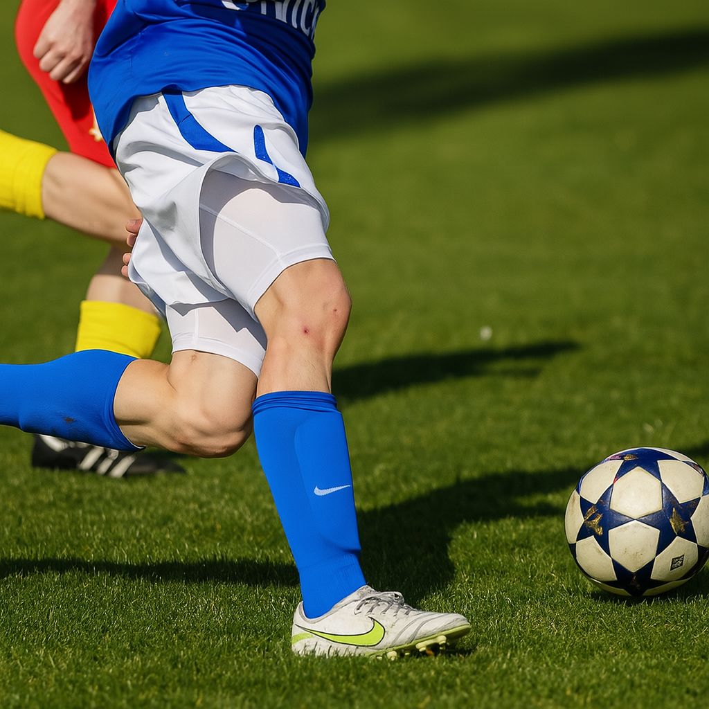
The development of cartilage damage is essentially possible due to two causes. In the case of accident-related cartilage damage, a piece of cartilage is sheared off from its surroundings as part of high force impacts (e.g. in the case of patellar dislocations). This is particularly common in sports injuries. Due to the enormous forces that occur in contact sports such as soccer, handball or football, there are also frequent accompanying injuries such as cruciate ligament or meniscus tears.
However, the articular cartilage is also subject to wear and tear over the course of life. This means that the texture of the cartilage tissue changes and there is therefore a higher susceptibility to damage. Therefore, long-term overloads (e.g. kneeling or squatting activities, leg axis misalignments or severe obesity) can lead to wear-related cartilage damage.
A special form of cartilage damage is osteochondrosis dissecans. This is a congenital circulatory disorder of the bone-cartilage junction. Frequently, after minor sports accidents (twisting, sprains), a bone-cartilage fragment is detached from the poorly perfused zone. This results in major damage and a free joint body which can cause blockages and further cartilage damage.
3. How is cartilage damage diagnosed?
The diagnosis of cartilage damage is usually made today by magnetic resonance imaging (MRI). Here, cartilage damage can already be diagnosed by an imaging procedure. In normal X-rays, the cartilage cannot be seen, only the bone structure. Another way to accurately diagnose cartilage damage is arthroscopy. Here, joint defects can be accurately described both in their extent and in their depth.
In principle, four degrees of severity of cartilage damage are described during arthroscopy. Previously, the classification was based on the so-called Outerbridge classification, today we recommend the internationally used ICRS classification:
- Grade I: intact surface with slight softening and minimal tears
- Grade II: abnormal cartilage with a lesion depth < 50% of the cartilage thickness
- Grade III: lesion depth > 50%, the cartilage defect extends to the bone layer depending on the gradation
- Grade IV: complete cartilage lesion, the bone is completely exposed – a so-called “cartilage bald spot”
4. What happens with untreated cartilage damage?
Since there are no blood vessels in the cartilage, the nutrients are introduced partly via the joint fluid and partly via the periosteum. Due to this poor nutrient supply, the hyaline articular cartilage cannot regenerate well. In the event of injury or wear of the cartilage structure, only an inferior cartilage structure is formed – a certain cartilage regeneration is only possible in children.
Cartilage damage to joints often leads to pain and progression of joint wear due to the lack of buffering and the occurrence of higher loads on the surrounding cartilage. Here, the mechanical load as well as the occurrence of the inflammatory reaction in the joint play a decisive role. If cartilage damage is not treated, it will inevitably increase due to the unfavorable force transmission at the edges of the damage.
At the end of cartilage damage is joint osteoarthritis. This is a degenerative disease of the joint, in which the damage is not limited to the cartilage and also affects the underlying bone. This leads to a narrowing of the joint space, to the compression of the bone tissue and to the formation of bony marginal attachments and cysts.
5. What role do combination injuries and pre-existing conditions play?
For the treatment of cartilage damage, the exact analysis of the accompanying injuries and diseases as well as risk factors of further damage is very important. If the cartilage damage is only considered in isolation, the therapeutic success is often at risk. For this purpose, an individual and unique diagnostic algorithm was developed in the MVZ Restart-Ortho.
In this context, accompanying injuries such as cruciate ligament injuries, patellar dislocations and meniscus damage are particularly important. If, for example, cartilage damage occurs as part of a patellar dislocation, stabilization of the patella is also crucial for long-term treatment success in addition to the therapy of the cartilage damage.
Also in the case of meniscus or cruciate ligament injuries, there is a disturbance of the mechanics of the knee joint and an instability which, if not corrected, prevents successful cartilage therapy. The same applies to misalignments of the leg axis. X or O-leg – misalignments lead to an overload of certain areas of the knee joint and can thus cause long-term overload-related damage.
In the case of misalignments of the leg axis, an exact analysis of the deformity is first carried out with the help of a so-called axis recording of the entire leg while standing. If it is then determined that a bony misalignment of the leg axis exists, it is possible to correct it with the help of an incision of the bone or the removal of a bone wedge. So that the bone does not close immediately, a small metal plate is attached, which stabilizes the bone until it has healed.
In the development of osteoarthritis from cartilage damage, concomitant diseases such as obesity and diabetes mellitus also play an essential role. These lead to a chronic inflammatory reaction, which changes the composition of the joint fluid and thus damages the articular cartilage.
6. Does cartilage damage always require surgery?
Since the cartilage tissue has no blood vessels, regeneration is only possible to a very limited extent. Non-operative treatment measures therefore aim to reduce pain, maintain the function of the joint and slow down the development of osteoarthritis. A repair of the cartilage is so far only possible surgically.
Through physical and physiotherapeutic measures, an optimization of the muscle guidance and an improvement of the mobility of the joint can be achieved. This allows muscular imbalances to be compensated. This leads to mechanical stabilization of the joint and also demonstrably has a positive influence on the inflammatory reaction.
In the case of high loads or axis misalignments, orthopedic measures can also be used to stabilize the joint. For example, the forces acting on the ankle or knee joint can be changed by special orthoses, both on the knee and on the ankle, as well as with appropriate insole care. This can also prevent cartilage damage from progressing, as a major cause of the development of this cartilage damage is the overload of certain joint sections.
A focus of the non-operative therapy of cartilage damage is the treatment of the inflammatory reaction. How exactly this occurs has not yet been fully understood. However, it is clear that small components of cartilage or meniscus tissue, which have been detached from their original environment, cause irritation of the synovial membrane.
For a long time, the classic, non-steroidal painkillers (ibuprofen, diclofenac) were the only treatment option to inhibit the inflammatory cascade in the joint. Nowadays, it is known that these should only be used for a short time due to their side effects.
So-called autologous blood procedures are playing an increasingly important role in the treatment of the inflammatory reaction. In this therapy, the body’s own mechanisms for regeneration are used. In human blood, in addition to the red and white blood cells, the platelets are dissolved. These contain hundreds of proteins, which play a central role in healing processes.
Another therapy option is the injection of hyaluronic acid. These improve the mechanical lubricity of the joint, because hyaluronic acid is a natural component of the joint fluid. There are studies that describe an improvement in the symptoms of affected patients, but the effectiveness seems to be limited in time.
Many patients try taking dietary supplements in addition to the therapy options described above. On the one hand, cartilage-protecting substances such as glucosamine and chondroitin should be mentioned, on the other hand, secondary plant substances such as curcuma and frankincense are increasingly advertised as anti-inflammatory. However, the scientific discussion about the effect of these substances has not been completed and there is still no reliable proof of efficacy at the present time.
7. What surgical treatment options are available?
Cartilage smoothing
Until the 1980s, cartilage smoothing (abrasion arthroplasty) was the standard method for treating cartilage damage. This involved surgically smoothing the damaged cartilage and removing bony growths. This procedure has since been abandoned due to poor results and should no longer be performed.
microfracturing
Microfracturing was developed in the 1980s. For many years, it was the standard procedure for treating cartilage damage. This procedure was first described by orthopedic surgeon Dr. R. Steadman. In principle, the defect is first cleaned of unstable cartilage fragments before several holes are drilled into the bone using a small drill or awl. The resulting bleeding causes tissue with cartilage-like properties to form. Studies have now shown that microfracturing is inferior to other cartilage therapy procedures in the long term. Therefore, microfracturing should now only be used for very small defects.
Matrix-associated bone marrow stimulation
Matrix-associated bone marrow stimulation was developed in the late 1990s as a further development of microfracturing. In this procedure, a membrane made of collagen or hyaluronic acid is sutured or glued to the defect after microfracturing to increase the stability of the blood clot. This so-called AMIC procedure has proven its superiority over microfracturing in studies and can be used on the knee and ankle for smaller cartilage defects (< 4.5 cm²).
Bone-cartilage transplantation
In cases of very small cartilage defects that also affect the bone, a bone-cartilage transplant is possible. This involves transplanting a cylinder from a less stressed joint area into the joint area where the cartilage damage is located and firmly inserting it there. In recent years, this has proven to be a good procedure for small defects. However, studies have shown that larger defects, which would require the removal of several cylinders, tend to have poor results.
Application of collagen gel
For very small defects, filling the defect with collagen gels has also become established practice. This does not require prior perforation of the bone. After the collagen gel is applied, cartilage cells from the surrounding cartilage are supposed to grow into the gel and stabilize it further. There aren’t any long-term study results for this procedure yet. However, this procedure isn’t suitable for larger cartilage defects.
Minced cartilage
In the minced cartilage procedure, cartilage tissue is first removed from the joint (for example, from the damaged area) and chopped into small cartilage chips. The minced cartilage is then applied to the defect and fixed in place with the help of blood platelets. This procedure was developed in the 1980s, but has become increasingly popular in recent years. Nevertheless, it must be emphasized that there are no major or long-term studies on this therapeutic procedure and it cannot therefore be recommended as standard for cartilage therapy.
autologous cartilage cell transplantation (ACT)
Cartilage cell transplantation has been used successfully for around 20 years. Numerous high-quality randomized studies have been conducted on this therapeutic procedure, which describe good results over a long-term period of more than 10 years. It is therefore the only cartilage therapy procedure recommended by the Working Group for Clinical Tissue Regeneration for the treatment of large cartilage defects (> 4.5 cm²). In matrix-associated autologous cartilage cell transplantation, cartilage cells are removed from a less stressed area of the knee joint during an initial operation and cultivated under highly sterile conditions in the laboratory over a period of several weeks. After a predefined period of 5-6 weeks, the new cartilage is inserted into the corresponding defect site. Although cartilage cell transplantation is expensive and time-consuming, it is currently the most promising procedure with the best long-term results.
Individual cartilage therapy
Thanks to our many years of experience, we are able to evaluate the many different cartilage regeneration procedures at our knee joint center on an individual basis to ensure the best possible recovery for each patient. After a diagnosis tailored to your symptoms, clinical picture, and physical demands, we will provide you with a personalized therapy recommendation. In addition to surgical treatment, there are numerous accompanying factors that can affect the outcome of the operation. At our center, we therefore place great emphasis on intensive follow-up treatment. First, the cartilage defect must be relieved of stress for several weeks. Usually, the operation is followed by customized physiotherapy and relief of the knee joint with the aid of an orthosis. The daily use of a motorized exercise splint (CPM) has also proven effective. As a rule, patients achieve normal range of motion after 2-3 months, so that the load can then be increased. However, contact sports should be avoided for a period of at least 12 months due to the higher shear forces acting on the cartilage.
8. What does the aftercare look like?
What should be considered during the first few days?
In the first few days after treatment for cartilage damage, the focus is on protecting the affected joint. It should be consistently cooled and elevated to reduce swelling. Passive movement may be prescribed by a doctor to maintain joint function. Stress and uncontrolled movements must be avoided at all costs.
Are crutches necessary?
Yes, walking aids such as forearm crutches are usually necessary to relieve the joint during the healing phase. The duration of relief depends on the procedure and the extent of the cartilage damage and is determined individually by the treating physician. Putting weight on the joint too early can jeopardize cartilage regeneration.
What physical exercises can be performed?
First, passive movement exercises are used, often with motorized continuous passive motion (CPM) devices, to promote joint mobility. This is followed by targeted physiotherapy exercises to strengthen the surrounding muscles and stabilize the joint. The exercises are gradually adapted and performed under professional guidance.
Aftercare schemes
PDF Aftercare cartilage damage