OPTIMAL TREATMENT OF MENISCUS INJURIES
The optimal treatment of meniscus injuries requires precise diagnostics and individually tailored therapy. The goal is to maintain joint function and sustainably alleviate discomfort.
- 1. What is a meniscus?
- 2. How does a meniscus injury occur?
- 3. What discomfort occurs after a meniscus injury?
- 4. How is a meniscus injury diagnosed?
- 5. What is the optimal therapy for meniscus injuries?
- 6. How is a meniscus surgery performed?
- 7. Frequently asked questions about meniscus surgery
- 8. Questions about treatment and aftercare regimens
1. What is a meniscus and what is its function?
The meniscus is a crescent-shaped cartilage structure in the knee joint that lies between the femur and tibia. Medial meniscus: It is located on the inside of the knee and is fused with the medial collateral ligament. This gives it less freedom of movement and makes it more susceptible to injury. Lateral meniscus: This is located on the outside of the knee and is more mobile, making it less likely to be injured.
The meniscus has several functions:
- Shock absorber: The meniscus distributes the load on the knee evenly to protect the articular cartilage by increasing the contact area between the femur and tibia, which reduces the pressure on the joint surfaces.
- Stabilization: It provides additional stability to the knee joint, especially during flexion and rotation movements.
- Joint lubrication and nutrition: They promote the distribution of synovial fluid, which supplies the cartilage with nutrients.
The menisci consist largely of fibrocartilage, which is very resilient. The outer edges of the menisci are supplied with blood, which facilitates regeneration there. The central areas, on the other hand, have no blood supply, which is why injuries in this area often heal poorly.
2. How does a meniscus injury occur?
A meniscus injury occurs when the meniscus is stressed beyond its load limit. This can happen due to sudden accidents or long-term wear and tear. There are two main causes of meniscus injuries:
Acute injuries:
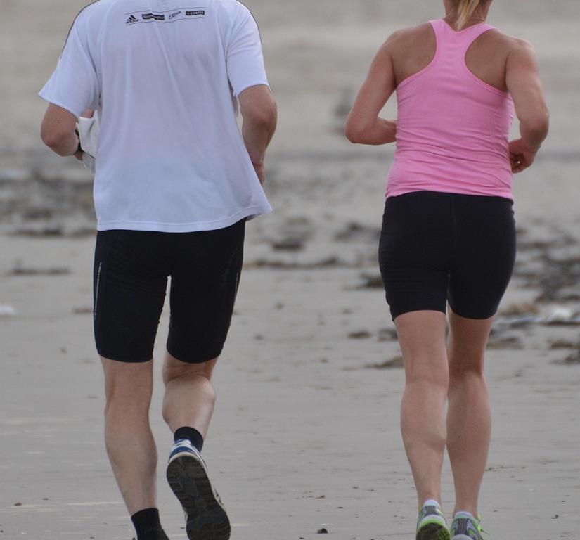
- Typical in sports: Sports such as soccer, skiing, or tennis, which require sudden turning or stopping movements, increase the risk.
- Mechanism: A meniscus tear often occurs when the femur is twisted against a fixed tibia.
Examples:
- A fall with a twisted knee.
- Abrupt turning movements, e.g. when changing direction in sports.
- Excessive strain, e.g. when lifting heavy loads in an unfavorable knee position.
Degenerative changes:
- Wear and tear due to age: As we age, the cartilage tissue in the meniscus becomes more brittle and less resilient.
- Chronic malalignment: Repeated stress, such as prolonged kneeling, squatting, or an unhealthy knee posture, leads to small damages that can worsen over time.
- Sudden symptoms with pre-existing damage: A seemingly harmless movement, such as standing up from a squat, can lead to a tear in a pre-damaged meniscus.
Meniscus tears can occur in different shapes, forms, and locations. The medial meniscus, which is fused to the collateral ligament, is usually affected. There are different types of meniscus tears that are medically well-defined, for example, longitudinal tears, transverse tears (radial tear), horizontal tears, and flap tears. Longitudinal tears occur as incomplete or complete tears. The bucket-handle tear is a longitudinal tear in the middle of the meniscus, in which the outer edges gape apart like a bucket handle.
A special but often overlooked form of meniscus injury is the root injury. Here, the acting forces cause the meniscus to tear out of its bony attachment. This severe injury interrupts the ring tension of the meniscus, and it can therefore no longer perform its function as a load distributor and shock absorber. If left untreated, root injuries therefore lead to rapidly progressive cartilage damage and the development of osteoarthritis.
4. Diagnosis of meniscus injury
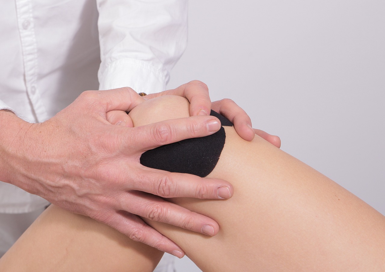
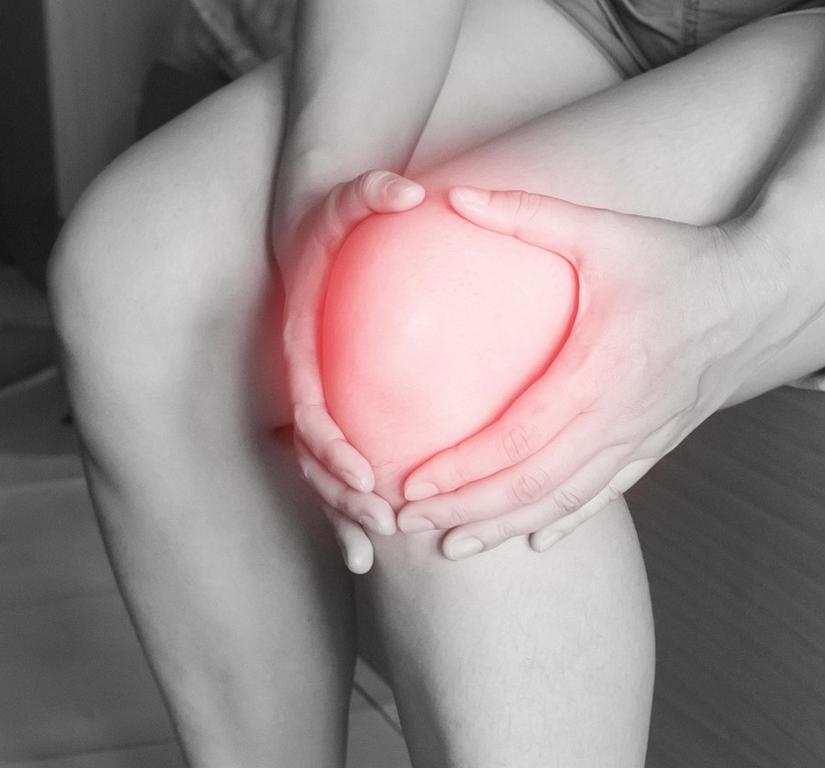
The diagnosis of a meniscus injury usually begins with a detailed discussion between the patient and the doctor. The accident, the type of symptoms, and any previous knee injuries are discussed. This is followed by a physical examination in which the doctor specifically checks the mobility of the knee joint and performs special tests to stress the meniscus and provoke painful movements or movement restrictions.
Typical, for example, is the pain when pressure is applied to the inner or outer joint space, which may indicate an injury to the medial or lateral meniscus. Rotating the lower leg with the knee bent is also tested to identify pain-sensitive areas. Such movement tests help to confirm the suspicion of a meniscus injury.
In order to confirm the diagnosis and rule out other knee injuries, imaging procedures are often used. Magnetic resonance imaging (MRI) is particularly important, as it allows the menisci, ligaments, and other structures in the knee joint to be shown in detail. An MRI is particularly helpful in identifying the exact location and extent of the damage. Conventional X-rays, on the other hand, only show the bony parts of the joint and are less helpful in assessing a meniscus injury, but can be used to rule out other problems such as osteoarthritis.
If no clear diagnosis is possible despite the imaging procedures, but the patient continues to have symptoms, arthroscopy (joint endoscopy) can be performed. A small camera is inserted into the knee joint to directly view the meniscus and other structures. Arthroscopy enables not only diagnosis but also immediate treatment, if necessary.
Through the combination of medical history, clinical examination, and modern imaging, a meniscus injury can usually be diagnosed precisely.
5. What is the optimal therapy for meniscus injuries?
The menisci are essential for protecting the articular cartilage. Therefore, the basic principle is that healthy meniscus tissue should be preserved under all circumstances and the repair of meniscus injuries should be promoted with all measures.
The optimal therapy depends not only on the type of tear but also on the patient’s age and concomitant injuries. Injury-related meniscus injuries in young patients should be operated on early, because if a meniscus injury remains untreated, it can lead to roughening of the articular cartilage or inflammation of the joint. A meniscus tear has the same effect as dirt in the gears of a machine: the longer torn pieces of tissue are left in the joint, the more damage can occur. An early diagnosis means an earlier start to treatment and, accordingly, less damage to the joint.
In the case of wear-related meniscus injuries in older patients, an indication for surgery should be made with caution, as the symptoms may also be caused by osteoarthritis. In these cases, physiotherapeutic treatment is initially indicated.
6. How is surgery performed on the meniscus?
The surgery on the meniscus is performed arthroscopically. The working instruments are inserted into the knee joint through two small incisions. The surgeon follows the procedure on a screen.
As part of the knee arthroscopy, an exact assessment of the meniscus injury with regard to its location and extent is possible. Depending on the type of meniscus injury, torn parts can be removed or reattached using suture techniques.
Fresh injuries, especially if they are located close to the base, i.e. close to the capsule, can be reattached with special techniques. Various suture techniques are available, which have been further developed in recent years and do not need to be removed again. If the bony attachment of the meniscus is torn off (meniscus root injury), surgery is usually necessary to fix the meniscus.
If a refixation of the torn meniscus is not possible, only the meniscus portion that is actually torn or unstable is removed. A complete or almost complete removal of the meniscus should be avoided at all costs.
7. Frequently asked questions about meniscus surgery
Is hospitalization necessary, or can the procedure be performed on an outpatient basis?
Meniscus surgery can usually be performed on an outpatient basis. After the operation, patients spend some time in the recovery room and are looked after by nursing staff. In the case of outpatient surgery, patients are taken home or picked up by relatives after the recovery period and follow-up care. A follow-up appointment for aftercare is scheduled at our practice the following day. If you have serious pre-existing conditions or are unable to receive care at home, meniscus surgery may also be performed on an inpatient basis under certain circumstances.
What type of anesthesia is required?
Die Gelenkspiegelung kann entweder in einer rückenmarksnahen Regionalanästhesie (Peridualanästhesie) – wobei von derArthroscopy can be performed either under regional anesthesia close to the spinal cord (epidural anesthesia), which numbs the area from the hips down, or under general anesthesia. Hüfte abwärts Schmerzfreiheit und Gefühllosigkeit erzeugt wird – oder in einer Allgemeinanästhesie (Vollnarkose) durchgeführt werden.
Are there any risks associated with meniscus surgery?
As with any surgical procedure, meniscus surgery carries a risk of inflammation and nerve and vascular injury, albeit to a lesser extent. Due to the partial immobilization of the leg after surgery, blood clots may form in the leg veins (thrombosis), with the risk of embolism. In rare cases, the operated meniscus may tear again.
8. Questions about treatment and aftercare regimens
What is the optimal follow-up treatment after meniscus surgery?
The optimal follow-up treatment after meniscus surgery includes controlled movement, targeted physical therapy, and a gradual increase in physical activity. The goal is to restore full joint function and avoid complications.
How long are crutches necessary?
The use of prescribed crutches varies depending on the surgical procedure. In the case of a partial meniscus resection, the crutches are used for approximately 3-4 days, during which time the operated leg may be placed on the ground. As a rule, the crutches can be dispensed with at home between the third and seventh day after the operation. They should continue to be used for longer distances, such as visits to the doctor. In most cases, the crutches no longer need to be used from the eighth postoperative day onwards. However, you should consult your treating physician. In the case of meniscus refixation, the crutches must be used for approx. 2 weeks to relieve the meniscus and ensure that it grows back securely.
What physical exercises can be performed?
The day after the operation, a specially tailored exercise program is started either at home or in the clinic. The aim of exercise therapy is to restore the mobility and resilience of the knee joint to the level it was at before the meniscus injury. This involves gradually increasing the range of motion, muscle strength, and flexibility of the upper and lower leg muscles. We pay particular attention to the extension of the knee joint, as this is the area where the risk of residual deficits is greatest. The intensity of the exercises and any restrictions on movement depend on the type of meniscus tear, the surgical procedure, and the goal of the therapy. For athletes, special movement exercises are performed toward the end of rehabilitation so that they can resume their sporting activities as soon as possible. The movement exercises after meniscus surgery serve to improve the mobility of the operated knee joint and strengthen the large thigh muscle.
Aftercare schemes
PDF aftercare partial meniscectomy
PDF aftercare meniscus refixation